In summary: Rhinoplasty is performed under general anesthesia in 3–4 hours and follows five distinct phases: preoperative assessment with clinical photographs, surgical marking in the upright position, anesthesia, the chosen approach (open or closed technique), and stepwise structural work on the dorsum and tip. The open technique adds a small 5–7mm transcolumellar incision for full direct visibility of the osteocartilaginous structure, making it preferred for complex corrections, asymmetries, and revision cases; the closed technique avoids external scars but offers more limited access. Cartilage for grafts is usually harvested from the nasal septum, with auricular cartilage used as a secondary source.
The question “how is rhinoplasty done” is the one most asked before a first consultation. It makes sense: it’s easier to decide on a surgical procedure when you know what’s going to happen in the operating room. This guide explains the real phases of the surgery, from preoperative assessment to the last suture.
Preoperative Assessment: Before Everything Starts
Rhinoplasty starts long before the operating room. The preoperative assessment is the moment when the surgeon analyzes your nasal anatomy in detail, defines the specific objectives of the procedure, and determines the most suitable technique for your case.
In that consultation, clinical photographs are taken in multiple positions — front, right and left profile, three-quarters, and base view — which serve both for surgical planning and postoperative comparison. If there’s a functional component (breathing difficulty), nasal exploration with a speculum or endoscopy is performed.
Preoperative tests are also performed: complete blood count, coagulation tests, electrocardiogram, and anesthetic evaluation. If the patient smokes, abstinence is required for at least 4 weeks before the scheduled date.
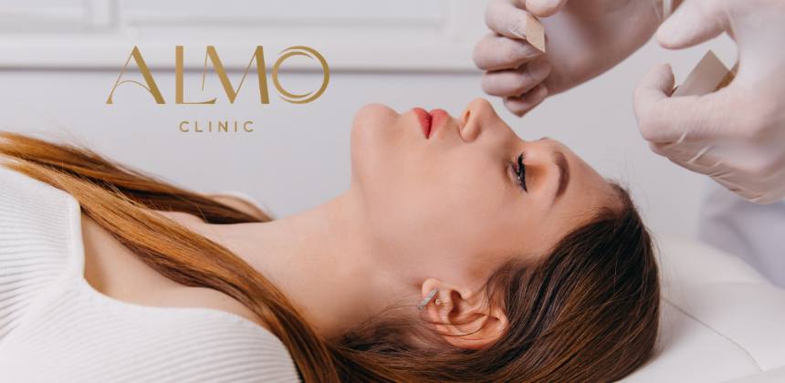
Preoperative Marking
On the day of surgery, in the preparation area, the surgeon makes the marking on the skin with the patient standing or sitting. He draws the incision lines, anatomical references, and work points. This marking in the upright position is fundamental because skin and soft tissues fall differently when the patient is lying down.
The marking defines exactly what will be removed, what will be repositioned, and what will be left intact.
Anesthesia: General or Deep Sedation
Rhinoplasty is performed under general anesthesia in most cases. The anesthesiologist administers anesthesia intravenously, followed by orotracheal intubation. The patient is completely unconscious and feels or remembers nothing from the procedure.
In some cases of minor rhinoplasty or exclusively tip surgery, deep sedation with local anesthesia supplementation may be used. However, for any correction involving bone work, general anesthesia is the safety standard.
Surgery lasts between 3 and 4 hours depending on the case complexity.
Approach: Open or Closed
The first step inside the operating room is the access incision. Here there are two main options:
Closed Technique (Endonasal)
All incisions are made inside the nasal cavities, in the internal mucosa. There is no visible external scar. Access is more limited than in the open technique, but sufficient for many corrections, especially those centered on the tip or cartilaginous dorsum without complex bone component.
The main advantage is the absence of external scar. The limitation is the surgeon’s lesser direct visibility of the structures to be modified.
Open Technique (External Approach)
A small transcolumellar incision (in the columella, the skin strip between the two nostrils) is added to the intranasal incisions. That incision, of about 5-7 mm, allows complete detachment of the skin from the nose and exposure of the entire osteocartilaginous structure with direct vision.
The open technique is most used in complex cases: rhinoplasties with extensive bone work, advanced tip corrections, revision cases, and significant asymmetries. The columellar scar is practically invisible at 6-12 months when the suture technique is correct.

Dorsal Nasal Work
Once the structure is exposed, the surgeon works in the order defined by the technique. In many cases, the dorsum is addressed first.
If there’s a dorsal hump, excess bone and cartilage forming that convexity is removed. The excess can be only cartilaginous (in the upper part of the septum, which is cartilage) or include bone (in the nasal bones forming the upper third of the dorsum). In bone corrections, the surgeon uses conventional instruments (chisel and rasp) or piezoelectric ultrasonic technique.
When reducing the dorsum, the nasal bones have an opening — an open roof — that must be closed through osteotomies: bone cuts on the sides of the nose that allow approximation of the bones toward the midline to close that opening and restore the nasal contour.
Tip Nasal Work
The nasal tip is the most complex part of rhinoplasty from a technical standpoint. It’s formed by two alar cartilages (the lateral lower cartilages) whose shape, position, and projection determine the tip’s appearance.
To refine a wide tip, the surgeon may suture the cartilages together, remove small portions of cartilage, or use special sutures that change the cartilages’ shape without removing tissue. To project a flat or dropped tip, a support graft (strut graft) or onlay cartilage graft may be used.
Cartilage needed for grafts usually comes from the nasal septum (the cartilage separating the two cavities). In cases where that cartilage is insufficient or has already been used, it may be taken from the auricular pavilion or, less frequently, from rib.
Septal Correction (When There’s Functional Component)
If the nasal septum is deviated and affects breathing, functional correction is performed in the same surgical time. The surgeon works inside the nasal cavities to straighten the deviated septum, removing or repositioning the portions obstructing air passage.
This part of the procedure transforms aesthetic rhinoplasty into septo-rhinoplasty, treating form and function simultaneously.
Closure and Splint Placement
Once corrections are completed, the surgeon closes the incisions with absorbable sutures inside and fine non-absorbable sutures outside (if open approach was used, removed between day 7 and 10).
An external splint and tapes are placed over the nasal dorsum to maintain the bones in their new position during consolidation. The splint remains in place between 8 and 20 days, depending on each case.
At ALMO we do not use nasal packing (as was done previously), so the patient leaves breathing well from the very first postoperative day.
Recovery Room and Discharge
The patient goes to the recovery room for 1 to 3 hours while anesthesia wears off completely. Most rhinoplasties are outpatient: the patient goes home the same day or with one night of hospitalization depending on complexity and clinic protocol.
Discharge includes written instructions for home care, scheduled analgesia, alarm signs, and date for the first postoperative check-up.
How Long Does Surgery Take
| Rhinoplasty Type | Approximate Duration |
|---|---|
| Tip Rhinoplasty (Closed) | 2 to 3 hours |
| Complete Rhinoplasty (Open) | 3 to 4 hours |
| Septo-Rhinoplasty | 3 to 4 hours |
| Secondary Rhinoplasty (Revision) | 3.5 to 4.5 hours |
Frequently Asked Questions
Does Rhinoplasty Hurt During Surgery?
No. You’re under general anesthesia and completely unconscious. You feel or remember nothing. Discomfort starts when anesthesia wears off in the first postoperative hours, and it’s controlled with oral analgesia.
What If I Wake Up During Surgery?
The risk of waking during general anesthesia exists statistically but is extremely rare with modern monitoring protocols. The anesthesiologist continuously monitors anesthesia depth throughout the surgery.
Can I See the Result Before Surgery?
The surgeon can do a photographic projection of the expected result during the assessment consultation. It’s an orienting reference, not a guarantee, because the final result depends on the specific characteristics of the patient’s skin and tissues.
Does Rhinoplasty Affect the Voice?
Not directly. If there’s septal correction with reduction of the nasal resonance space, there may be a slight change in voice resonance, especially if the patient has significant reduction rhinoplasty. In most cases, the change is minimal or imperceptible.
If you’re in the assessment phase, at ALMO Clinic the surgeon explains the entire procedure during consultation and resolves every doubt before defining any surgical date.